ORIGINAL RESEARCH | https://doi.org/10.5005/jp-journals-10024-2960 |
Reconstruction of Interimplant Papilla by Demineralized Freeze-dried Bone Allograft Block Fixed by Titanium Screw in Maxillary Esthetic Zone
1Department of Periodontology and Implantology, Sharad Pawar Dental College and Hospital, Wardha, Maharashtra, India
2Department of Periodontology, Vidarbha Youth Welfare Society’s Dental College, Amravati, Maharashtra, India
3Department of Periodontology and Implantology, Sharad Pawar Dental College and Hospital, Wardha, Maharashtra, India
4Department of Oral and Maxillofacial Surgery, Faculty of Stomatology, University of Belgrade, Serbia
5Department of Periodontology and Implantology, Chhatrapati Shahu Maharaj Shikshan Santha’s Dental College and Hospital, Aurangabad, Maharashtra, India
Corresponding Author: Kaustubh S Thakare, Department of Periodontology, Vidarbha Youth Welfare Society’s Dental College, Amravati, Maharashtra, India, Phone: +919890495485, e-mail: kaustubhthakaremds@gmail.com
How to cite this article Charde P, Thakare KS, Bhongade ML, et al. Reconstruction of Interimplant Papilla by Demineralized Freeze-dried Bone Allograft Block Fixed by Titanium Screw in Maxillary Esthetic Zone. J Contemp Dent Pract 2020;21(11):1205–1209.
Source of support: Nil
Conflict of interest: None
ABSTRACT
Aim: To evaluate effectiveness of demineralized freeze-dried bone allograft (DFDBA) block fixed by titanium screw for reconstructing interimplant papilla in maxillary esthetic zone during one-stage early loading multiple implant procedure.
Materials and methods: A total of 20 implants were placed in 10 systemically healthy patients (2 implants per patient) for replacement of multiple teeth by early loading one-stage implants along with interimplant papilla reconstruction using DFDBA block fixed by titanium screw. At the baseline, 6 months, and at 1 year, clinical measurements (interimplant papillary height measurement, papilla contour) and radiographic measurements were recorded.
Results: At 1 year, mean gain in interimplant vertical crestal bone was 1.7 mm, and complete reconstruction of the papilla was observed in 90% cases.
Conclusion: Demineralized freeze-dried bone allograft block fixed by titanium screw for reconstruction of interimplant papilla in maxillary esthetic zone during one-stage early loading multiple implant procedure is effective.
Clinical significance: Presence of interimplant papilla is of utmost importance for esthetically successful implant-supported restoration in the anterior region. This technique leads to reconstruction of interimplant papilla, thus providing esthetic appearance.
Keywords: Dental Implants, Interimplant, Papilla reconstruction.
INTRODUCTION
The oral rehabilitation of partially or fully edentulous patients can be done predictably by using dental implants.1–3
When multiple implants are used, the presence of interimplant papilla is very important for creating esthetically successful implant-supported restoration in anterior region of the oral cavity. The underlying osseous morphology has long been recognized as the foundation for the support of gingival tissue and interdental papilla. In a classic study, Tarnow et al. investigated the effects of crestal bone height on the presence or absence of dental papilla.4 The authors examined 288 interproximal sites and demonstrated that the papilla was present almost 100% of the time when the distance from the contact point to the crest of the bone was 5 mm or less. Salama et al. carried out a study suggesting a same correlation in implant therapy, in which the relationship between interproximal height of the bone (IHB) and papilla length was shown. Accordingly, IHB was classified into three classes:5 Class I IHB—4 to 5 mm (measured from bone crest to the apical extent of future contact point of the prosthesis) with predictable presence of papilla; Class II IHB—6 to 7 mm and guarded presence of papilla; and Class III—>7 mm IHB and in this class no papilla present. The data presented in the study demonstrate that the presence of papilla drops significantly and papilla cannot be recaptured, as the distance exceeds 5 mm in natural teeth and 3 mm in implants. Grunder et al. confirmed this speculation.6
Various soft tissue techniques have been attempted to reconstruct papilla. The soft tissue management techniques consist of free gingival grafts (Miller),7 coronally positioned flaps (Harvey),8 different types of pedicle grafts (Nelson),9 free connective tissue grafts, guided tissue regeneration procedures (Tinti et al.),10 and guided tissue augmentation (Salma et al.).11 However, based on the findings of Holmes, therapeutic techniques utilizing soft tissue management to reproduce interimplant papilla do not give predictable result.12
An interimplant papilla reconstruction attempt accomplishes predictable esthetics results when underlying labial and interproximal osseous support is provided.13 Till date, no study has reported hard tissue augmentation approach for the reconstruction of interimplant papilla, the present study was carried out with a basic aim of evaluating the effectiveness of demineralized freeze-dried bone allograft (DFDBA) block fixed by titanium screw for reconstructing interimplant papilla in maxillary esthetic zone during one-stage early loading multiple implant procedure.
MATERIALS AND METHODS
Ten healthy patients (7 males and 3 females) with a mean age of 26.2 ± 8.24 years (range 19–37 years) with multiple missing teeth in anterior area of maxillary jaw were selected from the outpatient department of periodontology and implantology, SP Dental College, Wardha, for placement of two implants in each patient. In total, 20 implants were placed in the study. Inclusion criteria were age ≥18 years, no pathology in edentulous area, sufficient bone volume at implant recipient site, D-1, D-2, or D-3 bone quality, and natural tooth with intact occlusal surface adjacent to edentulous space. Exclusion criteria were compromised general health jeopardizing the bone healing process, e.g., (diabetes, osteoporosis, blood disorders, and allergies to titanium), severe maxillomandibular space discrepancies, para functional habits, teeth showing spacing in interdental area/buccally inclined teeth/rotated or mal-aligned anterior teeth, history of alcoholism, excessive smoking or drug abuse, D-4 bone quality, and width of keratinized gingiva less than 2 mm at implant site.
Before starting the study, information concerning dietary status, oral hygiene maintenance habits, systemic background, gingival and periodontal status along with other routine clinical details were recorded in a specially designed chart. Patients were examined under good illumination with the help of mouth mirror and William’s graduated periodontal probe. Patients were made aware with the purpose and design of clinical study, and informed consent was signed from them. The ethical committee of Datta Meghe Institute of Medical Sciences, Wardha, approved the study protocol. Initial therapy consisting of oral hygiene instructions, supragingival and subgingival scaling, root planning under local anesthesia, and coronoplasty, if necessary, were performed after proper examination and diagnosis. Plaque control instructions were repeated until the patients achieved a plaque score of ≤1. Before entering the surgical phase, diagnostic cast of each patient was prepared to establish maxillomandibular relationship. A diagnostic wax up of the missing natural teeth and a clear acrylic resin surgical drill guide were prepared to facilitate correct implant placement. The clinical photograph, periapical and panoramic radiograph, and cone-beam computed tomography (CBCT) images to assess bone quality were obtained. Clinical and radiographic measurements recorded were:
Clinical Interimplant Papilla Measurement
Height of papilla (Grossberg criteria)14 and contour of papilla (Jemt index, 1997)15 were measured. These measurements were done at baseline, 6 months, and at 12 months after final restoration.
Papillary Height Measurement (Grossberg)14
Clinical photograph was obtained before surgery (Fig. 1) and at 6 months and 12 months postoperatively. On the photograph, a reference line was drawn from the most apical points of gingival margins on the adjacent teeth. Similar line was reproduced postoperatively. The most coronal extent of soft tissue to reference line distance was measured, and comparison of preoperative and postoperative photographs was used for calculating any variations in a vertical dimension of the soft tissue height.

Fig. 1: Preoperative clinical view showing missing 11, 12
Papilla Contour Measurement (Jemt 1997)15
It was measured by using papilla index score (PIS) from the clinical photograph obtained preoperatively and postoperatively at 6 months and 12 months after final restoration with score range 0–4. The index designates four different levels indicating the amount of papilla present. Assessment was measured from reference line drawn over the photograph through the highest gingival curvature on the adjacent permanent teeth on the buccal side. The distance from this line to contact point of the natural tooth/crown was assessed.
Radiographic Measurement of Interimplant Vertical Distance16,17
An intraoral periapical radiograph (IOPA) was taken at implant sites using long cone (XCP Rinn, Dentsply, New york, USA) paralleling technique at baseline, 6 months, and 12 months. A film mount having millimeter grid scale (Nix Company Ltd. Tokyo, Japan) was utilized for obtaining radiographic measurements. Vertical bone level at crest between implant (measured from the highest coronal point of bone crest to contact point after placement of prosthesis) was obtained by inserting the developed IOPA films in the mount.
In addition to measurement of interimplant vertical distance, bone quality assessment was done using CBCT. Hounsfield unit scale was used to assess the density of the bone, which in turn suggested quality of the bone. More than 600 HU in CBCT suggested very dense cortical bone, 400 and 600 HU suggested dense cortical-spongy bone, and less than 200 HU suggested cortical-spongy bone of low density.18
Implant Placement Using One-stage Early Loading Protocol
After induction of local anesthesia (xylocaine 2% solution comprising 1:100,000 epinephrine), using surgical blade No. 15, a horizontal palatal incision was placed 2 mm away from the crest of the ridge, care was taken not to split the adjacent papillae, and vertical releasing incision was then placed on labial surface extending to the vestibule. On labial and palatal side, full-thickness mucoperiosteal flap was raised. Ridge alveloplasty was done where needed. Surgical drill guide was used for placement of the pilot drill. Then, sequential drills were used to prepare implant site. Implants (Life care, Hi Tec one-stage implants) were placed in the recipient site (2 implants were placed in each patient). The neck of the implant was placed either at the crestal bone level or was kept slightly submerged. The abutment extension of the implant was kept such that the implant head protrudes about 2–3 mm from the bone crest.
Procedure for Papilla Reconstruction
Both the implants were placed at distance of ≥3 mm from each other. Midway between the two implants was drilled by a bur with a diameter 1 mm. Sterile saline solution was used to hydrate the bone block allograft (Freeze, dried, irradiated bone allograft block, provided by Tata Memorial Hospital, Tissue Bank, Mumbai, India) at least 45 minutes before use. A fissure bur in a high-speed hand piece with a copious saline solution was used to trim the block. Pre-drilling was performed on the prepared block to accommodate (1.5 mm × 8 mm) titanium screw (Orthomax Surgicals, Nagpur, India) (Fig. 2). Fixation screws were placed in a prepared block allograft obliquely to avoid possibility of stress fracture in the allograft. After stabilization of allograft block (Fig. 3), buccal flap was positioned around the implants and sutured to the palatal flap. After achieving complete tension-free closure, impressions were made and temporary acrylic resin crowns were cemented on the abutments. Providing immediate temporary prosthesis was helpful for guiding soft tissue. Temporary prosthesis was removed after 6 weeks, and a well-developed interimplant papilla was observed at this stage. Using high-viscosity vinyl polysiloxane final impression was made; a ceramic restoration was cemented onto the implants. The final prosthesis used was splinted.
Postoperative Care
All patients received (Cap. Amoxicillin, 500 mg t.i.d.) and (Tab. paracetamol 500 mg + ibuprofen 400 mg b. d.) postoperatively for 5 days. Patients were asked to avoid brushing teeth in the treated area and instead rinse three times per day with 0.2% chlorhexidine digluconate until suture removal. After 7–10 days of implantation, the sutures were removed.
Post-surgical Measurements
After giving permanent prosthesis, the patients were given appointments at 6 months and at 1 year. At every visit, clinical and radiographic measurements were done (Figs 4 and 5).
Statistical Analysis
Prior to initiating the study, statistical software power calculations (SPSS) were done. Results were calculated for clinical as well as radiographic parameters at baseline, 6 months, and at 12 months. Paired t test was utilized for assessing the statistical significance in relation to time points within the group for both clinical and radiographic measurements.
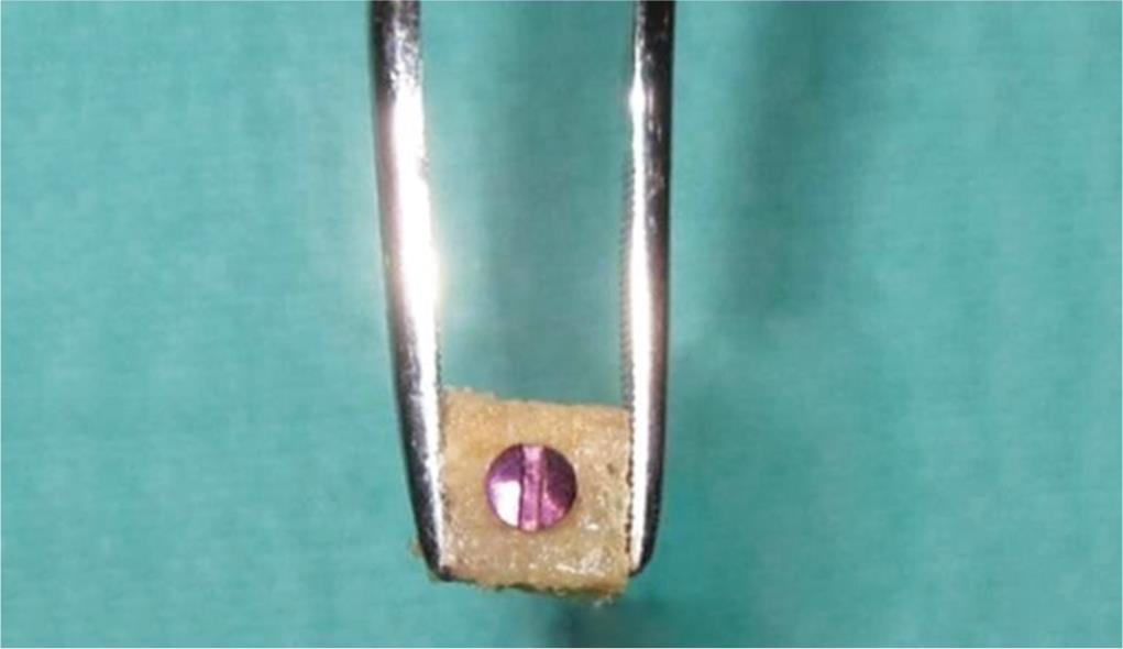
Fig. 2: Titanium screw placed in a prepared block allograft

Fig. 3: Stabilization of block with titanium screw in-between two implants

Fig. 4: Postoperative clinical view showing gain in interimplant papillary height

Fig. 5: Postoperative radiograph showing titanium screw between two implants
RESULTS
Clinical Measurements
Table 1 demonstrates variations in interimplant papillary height at baseline, 6 months, and 12 months. Table 2 shows percentage of papilla index scores at baseline, 6 months, and 12 months.
Radiographic Measurement
Table 3 shows radiographic interimplant vertical crestal bone gain at 6 and 12 months postoperatively.
DISCUSSION
In most of the cases when implant is used for replacing missing teeth in esthetic region of maxilla, buccal area and interproximal area of implants require augmentation of bone (Grunder et al.).19 Grossberg suggested that predictable esthetic results in implant therapy can be obtained by bone andsoft tissue augmentation techniques.14 Osseous supports when provided in the form of therapy can lead to predictable esthetic results in terms of reconstruction of papilla in interimplant area. In present study, DFDBA block which was fixed using titanium screw was utilized for guiding papilla to achieve a natural position of the papilla. At baseline, the mean height of papilla was 2 mm, and at 6 months this increased to 3.8 mm (increase of 1.8 mm). This height further increased to 4 mm at 12 months. Grossberg reconstructed papilla around implant with double pedicle flap at second-stage surgery, and 4 of 12 patients showed papilla formation with height of papilla up to 0.4–2.3 mm.14 Although the mean increase in height of papilla was not much, the author stated that a fixed reference line can be utilized for assessment of height of the papilla.
| At baseline (D1) | At 6 months (D2) | Gain in PH | At 1 year (D3) | Gain in PH |
|---|---|---|---|---|
| 2.00 ± 0.47 | 3.80 ± 0.63 | 1.80 ± 0.42 S | 4.00 ± 0.94 | 2.00 ± 0.81 S |
S, statistically significant (p < 0.05)
| S. no. | Months | Score-1 (esthetic failure) (%) | Score-2 (average esthetic) (%) | Score-3 (esthetic success) (%) |
|---|---|---|---|---|
| 1 | Baseline | 60 | 40 | 0 |
| 2 | 6 months | 0 | 50 | 50 |
| 3 | 1 year | 0 | 10 | 90 |
| At baseline | At 6-months | Vertical crestal bone gain at 6 months | At 1 year | Vertical crestal bone gain at 1 year |
|---|---|---|---|---|
| 6.60 ± 0.51 | 5.70 ± 0.48 | 0.90 ± 0.31 S | 4.90 ± 0.56 | 1.70 ± 0.67 S |
S, statistically significant (p < 0.05)
In our study, 90% of the cases showed (PIS-3) complete reconstruction of papilla, i.e. esthetic success, while 10% of cases showed (PIS-2) partially regenerated papilla, i.e., average esthetic at 12 months. This is explained by bone quality assessment; in 90% cases, the density of bone was very dense to dense cortical spongy bone, and these cases showed complete papilla regeneration. While one case showed cortical-spongy bone of low density and more marginal bone loss at 12 months, in this case the partial papilla regeneration occurred. Choquet analyzed crest of bone to contact point distance and its effect on papilla.13 When this distance was less than 5 mm, the presence of the papilla was seen 100% of the times. However, the presence of papilla was below 50% when this distance between crest of the bone and contact point was more than 5 mm. In our study, good esthetic outcome was observed in 9 cases (mean distance from crest of the bone to contact point was less than or equal to 5 mm), and in one case average esthetic success was seen, as the distance between the crest of the bone to contact point was 6 mm.
Mean gain of interimplant vertical crestal bone at 12 months was 1.7 mm. In a study by El-Salam El-Askary, three patients showed total regeneration of papilla by using insert of papilla made up of titanium and consisting of a core of titanium pyramidal in shape with height of around 2–3 mm and 3 mm base in buccolingual dimension and 1 mm in mesiodistal dimension.20 This study suggested that insert of papilla made up of titanium placed along with implants might help in establishing the papilla between two implants. Approach of guided regeneration of bone to create a bone peak in-between two implants has been proposed by Wohrle 2003,21 but bone regeneration depends on skills of clinicians and might be extremely unpredictable because of variation in resorption of bone. In the present study, trained doctors carried out all surgical procedures, and surgical approach used was one stage which could have resulted in limited marginal bone remodeling. Also in all the cases, emphasis was given to buccal bony plate and soft tissue preservation.
CONCLUSION
Over the entire observation period, soft tissue around implant was healthy, and 1.7 mm mean gain in interimplant vertical crestal bone was observed at 1 year. At 1 year reevaluation, complete reconstruction of the papilla was observed in 90% of cases.
LIMITATIONS
Following limitations may be noted in the present study:
- Long-term analysis is required to determine the stability of the results.
- Further well-controlled study is needed to confirm the findings of the present study with regard to interimplant papilla reconstruction.
CLINICAL SIGNIFICANCE
Successful implant-supported restoration when multiple implants are used in the maxillary anterior region of jaw basically is dependent on the papilla between the adjacent implants. Reconstruction of interimplant papilla in maxillary esthetic zone during one-stage early loading multiple implant procedure utilizing DFDBA block which is fixed with titanium screw can solve this problem and lead to a successful and esthetic implant-supported restoration.
REFERENCES
1. Branemark PI, Adell R, Breine U, et al. Intraosseous anchorage of dental prostheses. I. Experimental studies. Scan J Plastic Reconstr Surg 1969;3(2):81–100. DOI: 10.3109/02844316909036699.
2. Brånemark PI, Hansson BO, Adell R, et al. Osseointegrated implants in the treatment of the edentulous jaw. Experience from 10-year period. Scand J Plast Reconstr Surg Suppl 1977;16:1–132.
3. Schroeder A, Van der Zypen E, Stich H, et al. The reaction of bone, connective tissue and epithelium to endosteal implants with sprayed titanium surfaces. J Maxillofac Surg 1981;9:15–25. DOI: 10.1016/S0301-0503(81)80007-0.
4. Tarnow DP, Magner AW, Fletcher P. The effect of the distance from the contact point to the crest of bone on the presence or absence of the interproximal dental papilla. J Periodontol 1992;63(12):995–996. DOI: 10.1902/jop.1992.63.12.995.
5. Salama H, Salama MA, Garber D, et al. The interproximal height of bone: a guidepost to predictable aesthetic strategies and soft tissue contours in anterior tooth replacement. Pract Periodont Aesthet Dent 1998;10(9):1131–1141.
6. Grunder U. Stability of the mucosal topography around single-tooth implants and adjacent teeth: 1-year results. Int J Periodont Restorat Dent 2000;20(1):11–17.
7. Miller PDJr. Root coverage using a free soft tissue autograft following citric acid application: Part I. Technique. Int J Periodont Restorat Dent 1982;2:65–70.
8. Harvey PM. Management of advanced periodontitis: Part I. Preliminary report of method of surgical reconstruction. New Z Dent J 1965;61:180–187.
9. Nelson SW. The subpedicle connective tissue graft: a bilaminar reconstructive procedure for the coverage of denuded root surfaces. J Periodontal 1987;58(2):95–102. DOI: 10.1902/jop.1987.58.2.95.
10. Tinti C, Vincenzi G, Coltellini P, et al. Guided tissue regeneration in the treatment of human facial recession: a 12-case report. J Periodontol 1992;65(6):554–560. DOI: 10.1902/jop.1992.63.6.554.
11. Salma H, Salma M, Garber D, et al. Developing optimal periimpalnt papilla within esthetic zone: guided soft tissue augmentation. J Esthet Dent 1995;7(3):125–129. DOI: 10.1111/j.1708-8240.1995.tb00729.x.
12. Holmes CH. Morphology of the interdental papilla. J Periodontol 1965;36(6):21–26. DOI: 10.1902/jop.1965.36.6.455.
13. Choquet V, Hermans M, Adriaenssens P, et al. Clinical and radiographic evaluation of the papilla level adjacent to single-tooth dental implants. A retrospective study in the maxillary anterior region. J Periodontol 2001;72(10):1364–1371. DOI: 10.1902/jop.2001.72.10.1364.
14. Grossberg. D. Interimplant papilla reconstruction: assessment of soft tissue changes and results of 12 consecutive cases. J Periodontol 2001;72(7):958–962. DOI: 10.1902/jop.2001.72.7.958.
15. Jemt T. Regeneration of the gingival papillae after single- implant treatment. Int J Periodont Restorat Dent 2003;17:326–333.
16. Proussaefse P, Kan J, Lozada J, et al. Effects of immediate loading with threaded hydroxyapatite coated root-form implants on single premolar replacements: a preliminary report. Int J of Oral Maxillofac Implants 2002;17(4):567–572.
17. Turkyilmaz I, Avci M, Kuran S, et al. A 4-year prospective clinical and radiological study of maxillary dental implants supporting single-tooth crowns using early and delayed loading protocols. Clin Implant Dent Relat Res 2007;9(4):22–227. DOI: 10.1111/j.1708-8208.2007.00045.x.
18. Martinez H, Davarpanah M, Missika P, et al. Optimal implant stabilization in low density bone. Clin Oral Implants Res 2001;12(5):423–432. DOI: 10.1034/j.1600-0501.2001.120501.x.
19. Grunder U, Gracis S, Capelli M. Influence of the 3-D bone-to-implant relationship on esthetics. Int J Periodont Restorat Dent 2005;25:113–119.
20. El Askary AS. Use of a titanium papillary insert for the construction of interimplant papillae. Implant Dent 2000;9(4):358–362. DOI: 10.1097/00008505-200009040-00013.
21. Wohrle PS. Single-tooth replacement in the aesthetic zone with immediate provisionalization: fourteen consecutive case reports. Pract Periodont Aesthet Dent 1998;10:1107–1114.
________________________
© The Author(s). 2020 Open Access This article is distributed under the terms of the Creative Commons Attribution 4.0 International License (https://creativecommons.org/licenses/by-nc/4.0/), which permits unrestricted use, distribution, and non-commercial reproduction in any medium, provided you give appropriate credit to the original author(s) and the source, provide a link to the Creative Commons license, and indicate if changes were made. The Creative Commons Public Domain Dedication waiver (http://creativecommons.org/publicdomain/zero/1.0/) applies to the data made available in this article, unless otherwise stated.