

Probiotics in Caries Prevention
[Year:2018] [Month:February] [Volume:19] [Number:2] [Pages:2] [Pages No:123 - 124]
DOI: 10.5005/jp-journals-10024-2224 | Open Access | How to cite |
Abstract

Antifungal Activity of Xylitol against Candida albicans: An in vitro Study
[Year:2018] [Month:February] [Volume:19] [Number:2] [Pages:5] [Pages No:125 - 129]
DOI: 10.5005/jp-journals-10024-2225 | Open Access | How to cite |
Abstract
The most common fungal infection among human population is candidiasis, the etiology of which is mostly The standard strain of The MIC of xylitol for According to the results of this survey, xylitol has considerable antimicrobial effects. Hence, this substance can be used as an effective element in gums, toothpastes, and antimicrobial mouthwashes, especially in patients with candidiasis. By knowing the MIC and MFC of xylitol, this substance can be effectively used in products aimed to cure this fungal infection. Talattof Z, Azad A, Zahed M, Shahradnia N. Antifungal Activity of Xylitol against AimMaterials and methodsResultsConclusionClinical significanceHow to cite this article

[Year:2018] [Month:February] [Volume:19] [Number:2] [Pages:7] [Pages No:130 - 136]
DOI: 10.5005/jp-journals-10024-2226 | Open Access | How to cite |
Abstract
The aim of the study was to evaluate the effect of mouthrinses containing olive oil, fluoride, and their combination on enamel erosion. An Among the three mouthrinses, Listerine and Xerostom showed maximum protection against erosion on enamel. The 2% olive oil mouthrinse showed the least protection against erosion on enamel. The study indicated that the Listerine and Xerostom mouthrinses are valuable preventive measures. Mouthrinses are effective for lessening erosive demineralization and in aggregating remineralization of tooth surfaces which are the important factors to prevent enamel erosion. Vivek HP, Prashant GM, Geetha S, Chandramohan S, Imranulla M, Srinidhi PB. Effect of Mouthrinses containing Olive Oil, Fluoride, and Their Combination on Enamel Erosion: An AimMaterials and methodsResultsConclusionClinical significanceHow to cite this article

Effect of Green Tea Varnish on Depth of Root Caries
[Year:2018] [Month:February] [Volume:19] [Number:2] [Pages:6] [Pages No:137 - 142]
DOI: 10.5005/jp-journals-10024-2227 | Open Access | How to cite |
Abstract
Root caries is a common, debilitating condition particularly in the elderly population, which can lead to tooth loss. Evidence shows that green tea has cariostatic effects. Considering the gap of information on green tea varnish, this study aimed to assess the efficacy of green tea varnish in the prevention of root caries. This The mean (and standard deviation) depth of carious lesion was 54.30 ± 28.64 (μm), 0, and 0 in groups I, II, and III respectively. Control group showed the highest depth of caries. Groups II and III were not significantly different in this respect, but significant differences were noted in depth of caries between groups I and II and also groups I and III (p < 0.001). Based on the results, the green tea varnish may possess anticariogenic effects on root caries when applied every 24 or 48 hours over a 21-day period. Application of green tea varnish every 24 or 48 hours for 21 days may prevent root caries. Application of green tea varnish can prevent root caries. Daneshyar F, Khamverdi Z, Toliat T, Alikhani MY. Effect of Green Tea Varnish on Depth of Root Caries. J Contemp Dent Pract 2018;19(2):137-142.AimMaterials and methodsResultsConclusionClinical significanceHow to cite this article

[Year:2018] [Month:February] [Volume:19] [Number:2] [Pages:7] [Pages No:143 - 149]
DOI: 10.5005/jp-journals-10024-2228 | Open Access | How to cite |
Abstract
This Scanned digital images of 114 die preparations for all ceramic restorations (n = 114) were collected from a dental laboratory. All the images were also analyzed digitally using Adobe Photoshop® software to analyze the degree of taper (angle of convergence) of each preparation and then applied the Zuckerman's circle, and the Lewis perpendicular methods were used to measure the resistance form. For the current study, the overall average degree of taper was found to be 20.9° (range, 2—80°), which is more than what is recommended by most previous studies and also sharply greater than the textbook ideal of 3 to 6°. Mean degree of taper for maxillary was 17.56° (anterior—10.50°, posterior—23.7°), and for mandibular teeth, it was 25.22° (anterior—15°, posterior—28.45°). Out of the 64 analyzed images of maxillary teeth, 61 presented resistance form, while 3 were without it. Out of the 50 mandibular teeth analyzed, 38 possessed resistance form, whereas 12 were without. All the anterior teeth showed resistance form irrespective of the arch. The degree of taper showed a significant relationship with resistance and retention form, which was inversely proportional to each other. The recommended “degree of taper” is not always the clinically achievable as advocated in textbooks, as it is modified by various factors in the actual clinical situation. The study provides scientific background regarding the relationship between the degree of taper with resistance and retention form, and the relationship was found to be inversely proportional to each other. The recommended “degree of taper” is not always the clinically achievable as advocated in textbooks, and it is modified by various modifying or limiting factors in the actual clinical situation. Muruppel AM, Thomas J, Saratchandran S, Nair D, Gladstone S, Rajeev MM. Assessment of Retention and Resistance Form of Tooth Preparations for All Ceramic Restorations using Digital Imaging Technique. J Contemp Dent Pract 2018;19(2):143-149.AimMaterials and methodsResultsConclusionClinical significanceHow to cite this article

[Year:2018] [Month:February] [Volume:19] [Number:2] [Pages:6] [Pages No:150 - 155]
DOI: 10.5005/jp-journals-10024-2229 | Open Access | How to cite |
Abstract
The aim of the present study was to evaluate the effectiveness of different bristle designs of toothbrushes and the periodontal status among patients undergoing fixed orthodontic treatment. This randomized controlled trial (RCT) consisted of 45 adolescents (comprising 20 males and 25 females) undergoing fixed orthodontic treatment. The study participants were randomly allocated to three groups, each group being assigned a locally available toothbrush with a particular design of toothbrush bristle. In the first test phase, group I study participants were allocated to toothbrush with flat bristles, group II study subjects were allocated to toothbrush with zigzag bristles, and group III study participants were allocated to toothbrush with crisscross bristles. The study participants were recalled after 4 weeks to check the effectiveness of the allocated toothbrushes. A washout period of 1 week was maintained to ensure that there was no carryover effect of the different bristle designs. In the second test phase, each patient used the opposite toothbrush bristle design (group I: toothbrush with zigzag bristles, group II: toothbrush with crisscross bristles, and group III: toothbrush with flat bristles). Plaque scores were measured using Turesky—Gilmore—Glickman modification of Quigley-Hein plaque index (PI). In both phase 1 and 2 of this RCT, toothbrush with crisscross bristles exhibited maximum plaque reduction among the three different bristle design toothbrushes following 30 days (p = 0.312 ± 0.102 and 0.280 ± 0.110, respectively), which was statistically significant. It was concluded that all the three designs of toothbrushes were effective in removing plaque in patients with fixed orthodontic appliances. But among the three different toothbrushes, toothbrush with crisscross bristles showed the highest mean plaque reduction. Plaque accumulation around the orthodontic brackets and gingival margins is quite common among the fixed orthodontic patients, who encounter difficulty in maintaining good oral hygiene. Specially designed toothbrushes are very essential for effective plaque removal among the patients undergoing fixed orthodontic treatment. Naik SP, Punathil S, Shetty P, Jayanti I, Jalaluddin M, Avijeeta A. Effectiveness of Different Bristle Designs of Toothbrushes and Periodontal Status among Fixed Orthodontic Patients: A Double-blind Crossover Design. J Contemp Dent Pract 2018;19(2):150-155.AimMaterials and methodsResultsConclusionClinical significanceHow to cite this article

[Year:2018] [Month:February] [Volume:19] [Number:2] [Pages:10] [Pages No:156 - 165]
DOI: 10.5005/jp-journals-10024-2230 | Open Access | How to cite |
Abstract
The aim of this study was to evaluate the effect of different surface treatments on roughness, grain size, and phase transformation of presintered zirconia. Surface treatments included airborne particle abrasion (APA) before and after sintering with different particles shape, size, and pressure (50 μm Al2O3, 50 μm glass beads, and ceramic powder). Thirty-five square-shaped presintered yttrium-stabilized tetragonal zirconia polycrystal (Y-TZP) ceramic slabs (Zenostar ZR bridge, Wieland) were prepared (4 mm height × 10 mm width × 10 mm length) and polished with silicon carbide grit papers #800, 1000, 1200, 1500, and 2000 to ensure identical initial roughness. Specimens were divided into five groups according to surface treatment: group I (control): no surface treatment; group II: APA 50 μm Al2O3 after sintering; group III: APA 50 μm Al2O3 particles before sintering; group IV: APA 50 μm glass bead particles before sintering; and group V: APA ceramic powder before sintering. Specimens were analyzed using scanning electron microscopy (SEM), atomic force microscopy (AFM), X-ray diffraction (XRD) analyses, and tested for shear bond strength (SBS). Data were statistically analyzed using one-way analysis of variance (ANOVA) followed by Air abrasion before sintering significantly increased the surface roughness when compared with groups I and III. The highest tetragonal to monoclinic (t-m) phase transformation (0.07%) was observed in group III, and a reverse transformation was observed in presintered groups (0.01%). Regarding bond strength, there was a significant difference between APA procedures pre- and postsintering. Air abrasion before sintering is a valuable method for increasing surface roughness and SBS. The abrasive particles’ size and type used before sintering had a little effect on phase transformation. Air abrasion before sintering could be supposed to be an alternative surface treatment method to air abrasion after sintering. Skienhe H, Habchi R, Ounsi HF, Ferrari M, Salameh Z. Structural and Morphological Evaluation of Presintered Zirconia following Different Surface Treatments. J Contemp Dent Pract 2018;19(2):156-165.AimMaterials and methodsResultsConclusionClinical significanceHow to cite this article

[Year:2018] [Month:February] [Volume:19] [Number:2] [Pages:4] [Pages No:166 - 169]
DOI: 10.5005/jp-journals-10024-2231 | Open Access | How to cite |
Abstract
Both periodontitis and type II diabetes mellitus (T2DM) are common diseases with a multifactorial etiology and have influence of cytokines in their pathogenesis and thus may also influence each other. In recent times, more attention has been given to understanding the influences of these inflammatory cytokines which are a main part of oral chronic inflammation on systemic health of the individuals. Therefore, the aim of this study was to evaluate the plasma cytokine levels, specifically tumor necrosis factor-α (TNF-α), interleukin (IL)-6, and IL-4, in chronic periodontitis patients and T2DM patients, so as to investigate the influence of chronic periodontitis in systemic inflammation associated with diabetes mellitus. The present study comprised a total sample size of 60 patients. A detailed history along with complete periodontal examination were done for each person. These patients were subdivided into four study groups with 15 subjects (n = 15) in each group: group I: healthy individuals, group II: chronic periodontitis, group III: diabetes mellitus without chronic periodontitis, and group IV: diabetes mellitus with chronic periodontitis. Venous blood was withdrawn for obtaining serum samples from the subjects. Hemoglobin A1c (HbA1c) levels were measured from the automated chromatography. Using enzyme-linked immunosorbent assay kit, TNF-α, IL-4, and IL-6 were measured. It was observed that the difference between almost all the results showed statistical significance. Not much of a difference was seen when TNF-α and IL-6 findings of group II were compared with group III. Furthermore, IL-4 also did not differ when group II was compared with group IV. The inflammatory cytokines together control the inflammation process and a balance is maintained. However, in patients with diabetes mellitus, this balance is interrupted, which affects the final development and progression of the disease. Thus, hyperglycemia may be partly associated with the severity of the periodontal status in diabetic patients. Hyperglycemia thus may play a role in increasing the severity of the periodontal status in diabetic patients. Keeping such relationship in mind, better treatment modalities can be provided to the patients. Bakshi D, Kaur G, Singh D, Sahota J, Thakur A, Grover S. Estimation of Plasma Levels of Tumor Necrosis Factor-α, Interleukin-4 and 6 in Patients with Chronic Periodontitis and Type II Diabetes Mellitus. J Contemp Dent Pract 2018;19(2):166-169.IntroductionMaterials and methodsResultsConclusionClinical significanceHow to cite this article

[Year:2018] [Month:February] [Volume:19] [Number:2] [Pages:7] [Pages No:170 - 176]
DOI: 10.5005/jp-journals-10024-2232 | Open Access | How to cite |
Abstract
The aim of the study was to evaluate the proliferative index (PI) at their invasive front of oral cancer and their association with Bryne's grades of oral squamous cell carcinoma (OSCC) and compare the PI with lymph node metastasis, site of involvement, and habits. The Ki-67 antigen expression was immunohistochemically evaluated in a total of 102 cases that included the histopathologically diagnosed archival specimens of OSCCs. They were subdivided by Bryne's histopathological grading into grade I (40 cases), grade II (32 cases), and grade III (30 cases). The nucleus with brown stain was considered positive. Cells were counted under 400× magnification. The proliferative activity thus determined was then expressed as a percentage of Ki-67 labeling index (Ki-67 LI) positive cells. A stepwise increase in the mean Ki-67 LI was found from grade I to III squamous cell carcinoma, thus correlating with the histological grading. In addition, there was a higher PI seen in cases associated with metastatic lymph node, which concords with the higher biologic aggressiveness and poor prognosis of the lesion. The present study shows a definitive correlation of Ki-67 antigen with the Bryne's histological grading, all the parameters of Bryne's grading for OSCC and lymph node status of the patient proving its association as an effective tool to grade the tumors and finally read the prognosis of the tumor. Cell proliferation is regarded as one of the most important biologic mechanisms in oncogenesis. The role of cell proliferation in tumor progression has been inferred in studies concerned with human cancer by comparing the PI of normal tissue, preneoplastic and neoplastic lesions. The Ki-67 antigen-labeled cells can prove to be an effective aid to grade the tumors. It might be possible to standardize and objectify tumor grading among pathology laboratories. Bhuyan L, Sarangi S, Das BK, Das SN, Nayak S. Proliferative Index in Invasive Tumor Front of Oral Squamous Cell Carcinoma: A Potential Prognostic Indicator. J Contemp Dent Pract 2018;19(2):170-176.AimMaterials and methodsResultsConclusionClinical significanceHow to cite this article

Spirometric Assessment of Impact of Complete Dentures on Respiratory Performance: An in vitro Study
[Year:2018] [Month:February] [Volume:19] [Number:2] [Pages:4] [Pages No:177 - 180]
DOI: 10.5005/jp-journals-10024-2233 | Open Access | How to cite |
Abstract
Edentulism is one of the debilitating states affecting a significant portion of the geriatric population. It is often considered as the ultimate marker for predicting the pressure and burden on oral health. Respiration is also a routine vital function running continuously through the human body and most commonly assessed by a spirometer. Hence, we planned the present study to assess the impact of complete dentures on the functioning of the respiratory system. In the present study, a spirometric assessment of the effect of complete dentures on respiratory performance was done. A total of 100 subjects were included, and diagnostic spirometer was used for carrying out the spirometric test at different stages of each subject. The spirometric test was carried out at four different stages: In the absence of both the denture (AODs), with both the dentures (maxillary and mandibular) inserted in the patient's mouth (BDs), with only maxillary denture inserted in the oral cavity (UDs), and finally, by inserting only the mandibular dentures in the oral cavity (LDs). Forced vital capacity (FVC), peak expiratory flow (PEF), forced expiratory volume in 1 second (FEV1), and forced expiratory flow between 25 and 75% (FEF25—75) were evaluated. All the results were compiled and assessed using Statistical Package for the Social Sciences (SPSS) software. Of the total 100 subjects included in the study, 42 were males and 58 were females. The mean FVC values of AOD, BD, LD, and UD group were 3.10, 3.02, 2.90, and 2.93 respectively. The mean PEF values of AOD, BD, LD, and UD group were 5.79, 5.60, 5.40, and 5.48 respectively; 2.39, 2.35, 2.33, and 2.32 were the mean FEV1 values observed in AOD, BD, LD, and UD group respectively. Statistically significant results were obtained while comparing AOD—FVC and BD—FVC and other oral conditions. Spirometric values of respiratory functional tests in edentulous patients might be unfavorably affected by wearing complete dentures. Since subjects without complete dentures exhibited a maximum value of respiratory functional test, respiratory exercise protocols should also be carried in denture-wearing edentulous patients for increasing the performance of the respiratory system. Indrakumar HS, Venkatesh D, Adoni VV, Kashyap R, Jayanthi D, Prakash N. Spirometric Assessment of Impact of Complete Dentures on Respiratory Performance: An IntroductionMaterials and methodsResultsConclusionClinical significanceHow to cite this article

Surface Characterization and Cell Adhesion of Different Zirconia Treatments: An in vitro Study
[Year:2018] [Month:February] [Volume:19] [Number:2] [Pages:8] [Pages No:181 - 188]
DOI: 10.5005/jp-journals-10024-2234 | Open Access | How to cite |
Abstract
The aim of this study was to characterize the surface of zirconia subjected to different treatments and evaluate its effect on cell adhesion and proliferation. A total of 80 zirconia disks were divided into four groups (n = 20) according to the surface treatments used: group I: as-sintered (AS), no surface treatment applied; group II: abrasion treatment applied using Rocatec (ROC; 3M ESPE) system with silica-coated alumina powder of grit size 110 μm; group III: erbium, chromium:yttrium, scandium, gallium, garnet (Er, Cr:YSGG) laser (LAS; BIOLASE) was used at a frequency of 20 Hz and output power of 3 W; and group IV: specimens were subjected to the selective infiltration etching (SIE) technique. Surface characterization was evaluated for the different groups (roughness, hardness, and morphology), and cell behavior (adhesion and proliferation) was tested (α = 0.05). The ROC group reported a significant increase in surface roughness (2.201 ± 0.352) and Vickers hardness (1758 ± 16.6) compared with the other surface treatments. The SIE surface-treated group reported a significantly higher number of cells (64.5 ± 2.6 and 53.5 ± 2.2 respectively) compared with the other surface-treated groups. The SIE is a promising surface treatment for zirconia that significantly enhances cell adhesion and osseointegration. The SIE treatment of zirconia implants may help in a faster and better osseointegration. Nassif W, Rifai M. Surface Characterization and Cell Adhesion of Different Zirconia Treatments: An AimMaterials and methodsResultsConclusionClinical significanceHow to cite this article
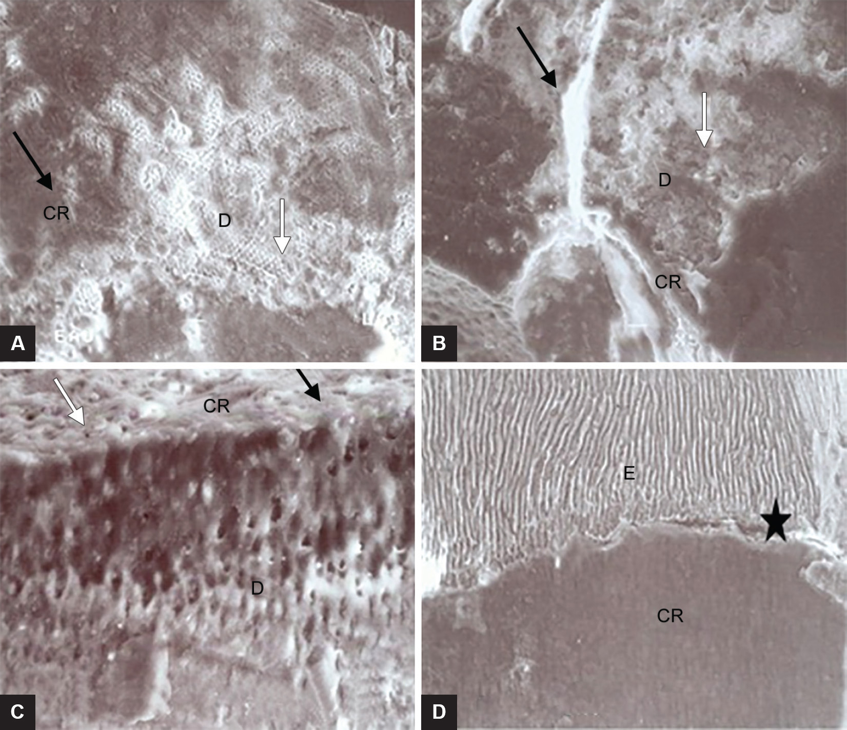
Microtensile Bond Strength of Polyacid-modified Composite Resin to Irradiated Primary Molars
[Year:2018] [Month:February] [Volume:19] [Number:2] [Pages:7] [Pages No:189 - 195]
DOI: 10.5005/jp-journals-10024-2235 | Open Access | How to cite |
Abstract
This study evaluated the influence of various doses of radiotherapy on the microtensile bond strength (μTBS) of compomer resin to dentin and enamel in primary molars. Thirty-five intact primary molars were collected and divided into seven groups. Teeth were irradiated with doses from 10 to 60 Gy, except for the control group. Compomer restorations were performed, and enamel—compomer resin beams and dentin—compomer resin beams were tested at a crosshead speed of 1 mm/min. No statistically significant difference was found between the irradiated tooth enamel and the control group ( Radiation may not cause a significant difference in the μTBS of compomer resin to primary tooth enamel, but appears to dose dependently decrease its bond strength to primary tooth dentin. Radiotherapy may affect the success rate of compomer fillings in primary teeth, especially in deeper cavities with exposed dentin. Keles S, Yilmaz Y, Sezen O. Microtensile Bond Strength of Polyacid-modified Composite Resin to Irradiated Primary Molars. J Contemp Dent Pract 2018;19(2):189-195.AimMaterials and methodsResultsConclusionClinical significanceHow to cite this article

[Year:2018] [Month:February] [Volume:19] [Number:2] [Pages:9] [Pages No:196 - 204]
DOI: 10.5005/jp-journals-10024-2236 | Open Access | How to cite |
Abstract
This study evaluated the effect of light-emitting diode (LED) illumination bleaching technique on the surface nanohardness of various computer-aided design and computer-aided manufacturing (CAD/CAM) ceramic materials. Twenty disk-shaped samples (width, length, and thickness = 10, 15, and 2 mm) were prepared from each of the ceramic materials for CAD/CAM, including Lava™ Ultimate (LV), Vita Enamic® (En) IPS e.max® CAD (Me), inCoris® TZI (IC), and Prettau® zirconia (Pr). The samples from each type of ceramic were randomly divided into two groups based on the different bleaching techniques to be used on them, using 35% hydrogen peroxide with and without LED illumination. The ceramic disk samples were bleached according to the manufacturer's instruction. Surface hardness test was performed before and after bleaching using nanohardness tester with a Berkovich diamond indenter. The respective Vickers hardness number upon no bleaching and bleaching without or with LED illumination [mean ± standard deviation (SD)] for each type of ceramic were as follows: 102.52 ± 2.09, 101.04 ± 1.18, and 98.17 ± 1.15 for LV groups; 274.96 ± 5.41, 271.29 ± 5.94, and 268.20 ± 7.02 for En groups; 640.74 ± 31.02, 631.70 ± 22.38, and 582.32 ± 33.88 for Me groups; 1,442.09 ± 35.07, 1,431.32 ± 28.80, and 1,336.51 ± 34.03 for IC groups; and 1,383.82 ± 33.87, 1,343.51 ± 38.75, and 1,295.96 ± 31.29 for Pr groups. The results indicated surface hardness reduction following the bleaching procedure of varying degrees for different ceramic materials. Analysis of variance (ANOVA) revealed a significant reduction in surface hardness due to the effect of bleaching technique, ceramic material, and the interaction between bleaching technique and ceramic material (p < 0.05). Bleaching resulted in a diminution of the surface hardness of dental ceramic for CAD/CAM. Using 35% hydrogen peroxide bleaching agent with LED illumination exhibited more reduction in surface hardness of dental ceramic than what was observed without LED illumination. Clinicians should consider protection of the existing restoration while bleaching. Juntavee N, Juntavee A, Saensutthawijit P. Influences of Light-emitting Diode Illumination Bleaching Technique on Nanohardness of Computer-aided Design and Computer-aided Manufacturing Ceramic Restorative Materials. J Contemp Dent Pract 2018;19(2):196-204.AimMaterials and methodsResultsConclusionClinical significanceHow to cite this article

[Year:2018] [Month:February] [Volume:19] [Number:2] [Pages:5] [Pages No:205 - 209]
DOI: 10.5005/jp-journals-10024-2237 | Open Access | How to cite |
Abstract
The aim of this study is to perform three-point bend test on submicron hybrid composite fabricated with direct and indirect veneer technique. A total of 20 maxillary anterior teeth were selected, and labial reduction of 0.5 to 0.75 mm with a chamfered finish line for veneer preparation was done. Teeth were divided into two groups depending on fabrication technique being used: group I—veneers fabricated with light and group II—veneers fabricated with light and heat (PHOTOPOL). Specimens were tested under universal testing machine (UTM) where load was applied at a crosshead speed of 1 mm/min with a pointer of 1 mm diameter. Data were statistically analyzed. The results showed highly significant difference between the two groups with the mean value of group I (246.7 ± 2.285 N) and group II (531.1 ± 4.411 N). The curing mechanism involving light and heat increases the fracture resistance of the veneers. Within the limitations of this study, the results led to the conclusion that the association of common composites with a simple postcure heat treatment may be an alternative for current indirect composite systems, although more studies are needed to assess other properties of the composites for this application. Sethi A, Makkar S, Sidharth K, Kaur T, Sandhu SS, Joseph AK. Evaluation of the Flexural Strength of Submicron Hybrid Composite using Different Fabrication Methods: An AimMaterials and methodsResultsConclusionClinical significanceHow to cite this article

[Year:2018] [Month:February] [Volume:19] [Number:2] [Pages:4] [Pages No:210 - 213]
DOI: 10.5005/jp-journals-10024-2238 | Open Access | How to cite |
Abstract
The aim of this study is to find if there is any correlation between the hematological parameters and temporomandibular joint (TMJ) ankylosis and severity of the disease in such patients when compared with the nonankylosed patients. A total of 70 patients with age ranging from 10 to 40 years were included in the study after excluding the subjects according to the inclusion criteria. We categorized the subjects into two major groups: group I: control (nonankylosed/healthy subjects) and group II: study group (ankylosed subjects) with each group containing 35 subjects (n = 35) respectively. A detailed personal and medical history was obtained. The pharynx diameter was also recorded for each patient, and blood investigations using venous blood were done, which included hemoglobin concentration and hematocrit values. The results of study population showed a mean age of 22 ± 2.2 years. The most common etiology reported was trauma (65.7%) followed by infections, in which Noma was the most common one (80%). The difference of the mean values for hemoglobin and hematocrit concentration, between both the groups, was found to be statistically significant (p < 0.0001). Furthermore, a positive correlation was observed between the hemoglobin concentration and duration of ankylosis. This study was an attempt to find a relation between the hemoglobin and hematocrit values in TMJ ankylosis patients so that the clinical treatment and management of such patients during surgeries be improved and may be beneficial for the patient. Temporomandibular joint ankylosis patients have to undergo complex surgical treatment, where the risk of excessive blood loss is high. Therefore, considering the complications of blood transfusions, such as infections and other risk factors, these patients can be good subjects for autologous blood transfusions, which help in improvement of the overall well-being of the patient. Singh D, Kaur G, Bakshi D, Sahota J, Thakur A, Grover S. Evaluation of Hemoglobin Concentration and Hematocrit Values in Temporomandibular Joint Ankylosis Patients in Comparison to Nonankylosed Patients. J Contemp Dent Pract 2018;19(2):210-213.AimMaterials and methodsResultsConclusionClinical significanceHow to cite this article

[Year:2018] [Month:February] [Volume:19] [Number:2] [Pages:7] [Pages No:214 - 220]
DOI: 10.5005/jp-journals-10024-2239 | Open Access | How to cite |
Abstract
Intraoperative antibiotics may be effective in elective surgery; there may be an advantage to starting antibiotics preoperatively when there is already an infective focus, such as compound facial fractures. The purpose of this study is to compare preoperative and intraoperative antibiotic prophylaxis in compound facial fractures. This is a prospective study conducted over a period of 2 years on 50 patients, who underwent open reduction and rigid internal fixation. The patients were assigned to two groups. The patients in group I received antibiotics at the time of admission. The patients in group II received antibiotics perioperatively at the time of induction of general anesthesia. Postoperatively, the patients were evaluated for the reduction of pain and the presence of infection by assessing the local presence of erythema, swelling, a rise in temperature, and purulent discharge, if any, on a predefined proforma. A total of 72 fractures were assessed in 50 patients. There was an overall reduction of pain in both the groups. Wound healing status was found to be satisfactory in both the groups. Wound status and infection rate were evaluated in patients of both the groups. In the study, there was no difference between the two groups on the predecided parameters whether the antibiotics were given either preoperatively or perioperatively. It is very essential to have a sound knowledge of the use of antibiotic therapy effectively to prevent the overuse of it and, thereby, help avoid developing resistance to antibiotics in patients. Mamthashri V, Reddy BP. Comparison of Preoperative and Perioperative Antibiotic Prophylaxis Regimen in Compound Facial Fractures. J Contemp Dent Pract 2018;19(2):214-220.Aims and objectivesMaterials and methodsResultsConclusionClinical significanceHow to cite this article

Effect of Bleaching Mouthwash on Force Decay of Orthodontic Elastomeric Chains
[Year:2018] [Month:February] [Volume:19] [Number:2] [Pages:5] [Pages No:221 - 225]
DOI: 10.5005/jp-journals-10024-2240 | Open Access | How to cite |
Abstract
Force decay elastomeric chains are significant, and it is a clinical problem. The aim of this study was to evaluate the effects of bleaching agent in the mouthwash on the force decay of orthodontic chains. In this experimental study, 160 gray closed elastomeric chains were randomly divided into three groups (one control and two test groups). Four loops of chains were stretched for 25 mm on custom-made jig. Control group specimens were immersed in artificial saliva during the test period. Test group specimens were immersed twice a day for 30 seconds in the whitening (LISTERINE® HEALTHY WHITE™) and daily sodium fluoride (LISTERINE® TOTAL CARE ZERO) mouthwashes. All specimens were immersed in artificial saliva at 37°C. Force was measured at different time points (initial, 1, 7, 14, 21, 28 days). Statistical analysis was performed by two-way analysis of variance (ANOVA) and Bonferroni methods ( Force of elastomeric chains was decreased dramatically in all groups during the experiment. After 24 hours, force was decreased by 42.18, 48.34, and 53.38% in control group, daily, and bleaching mouthwash groups respectively. The corresponding numbers after 4 weeks were 66.30, 76.73, and 86.48. The difference between three groups at days 1 and 28 was statistically significant (p < 0.05). Within the limitations of the current Use of whitening mouthwash by orthodontic patients could decrease the force of elastomeric chains, so it could be recommended to use them for a short time. Behnaz M, Namvar F, Sohrabi S, Parishanian M. Effect of Bleaching Mouthwash on Force Decay of Orthodontic Elastomeric Chains. J Contemp Dent Pract 2018;19(2):221-225.IntroductionAimMaterials and methodsResultsConclusionClinical significanceHow to cite this article

[Year:2018] [Month:February] [Volume:19] [Number:2] [Pages:7] [Pages No:226 - 232]
DOI: 10.5005/jp-journals-10024-2241 | Open Access | How to cite |
Abstract
The aim of this study was to clinically compare glass ionomer cement (GIC) with microhybrid composite resin used in class I cavities on permanent teeth over a period of 9 months. A total of 40 teeth with class I cavities were divided into two groups (n = 20) and restored with GIC (EQUIA; GC) and microhybrid resin composite (Amelogen Plus; Ultradent). Restorations were evaluated at ×4.5 magnification using the United States Public Health Service (USPHS) criteria every 3 months. Statistical analysis was performed using the Fisher's exact test (α < 0.05). The data obtained reported no statistical significance difference between both groups in regard to anatomical shape, color, postoperative sensitivity, secondary caries, material handling, adaptation, and marginal staining. The results of this clinical study showed that GIC (EQUIA; GC) can be used for the restoration of permanent teeth and may be more appropriate for certain clinical situations than the resin composite material. EQUIA (GIC) is a viable alternative to resin composite in restoring class I cavities in permanent teeth. Kharma K, Zogheib T, Bhandi S, Mehanna C. Clinical Evaluation of Microhybrid Composite and Glass Ionomer Restorative Material in Permanent Teeth. J Contemp Dent Pract 2018;19(2):226-232.AimMaterials and methodsResultsConclusionClinical significanceHow to cite this article

Middle Mesial and Middle Distal Canals in Mandibular First Molar
[Year:2018] [Month:February] [Volume:19] [Number:2] [Pages:4] [Pages No:233 - 236]
DOI: 10.5005/jp-journals-10024-2242 | Open Access | How to cite |
Abstract
Root canal anatomy is a complex entity. The main objective of root canal treatment is to get rid of the infection and have a good apical and coronal seal with an appropriate filling. Inability to achieve thorough cleaning and shaping followed by three-dimensional obturation of the root canal system usually causes root canal treatment failure. For this reason, clinicians should be aware of these anatomical variations to achieve successful treatment. The aim of this article is to report on the successful treatment and follow-up of mandibular first molar with additional middle mesial (MM) and middle distal (MD) canals. A 29-year-old white male patient reported with a complaint of pain in relation with tooth #19. On clinical examination, diagnosis of symptomatic irreversible pulpitis with symptomatic apical periodontitis and condensing osteitis was made and nonsurgical root canal treatment was planned. Initially, two mesial and two distal canals were located, and the patient was planned for the obturation in the second visit. The complaint of mild persistent symptoms gave a possibility of additional canals. Under the dental operating microscope and selective troughing on the floor of the pulp chamber with ultrasonic tips, additional canals were located as MM and MD canals. Leaving some area of the root canal system untreated is found to be one of the main reasons for root canal treatment failure. Dentists should take advantage of new tools, such as dental operating microscope and ultrasonic tips to be able to locate and treat the hidden and unusual anatomy. Mandibular first molar with six canals is very rare to encounter. Clinician should have a thorough knowledge of these unusual anatomy to avoid treatment failure due to incomplete disinfection of the root canal system. Additional canals, Mandibular first molar, Middle distal, Middle mesial, Root canal morphology. Jabali AH. Middle Mesial and Middle Distal Canals in Mandibular First Molar. J Contemp Dent Pract 2018;19(2):233-236.BackgroundAimCase reportConclusionClinical significanceKeywordsHow to cite this article

[Year:2018] [Month:February] [Volume:19] [Number:2] [Pages:5] [Pages No:237 - 241]
DOI: 10.5005/jp-journals-10024-2243 | Open Access | How to cite |
Abstract
Parental presence/absence in the dental operatory (also called: Parent-in—parent-out technique) is an extremely controversial aspect of the nonpharmacological BMTs. Historically, dentists used to exclude parents from dental operatory to avoid their interference with the dentist's aptitude to build a rapport and relationship with the child, hence increasing the child management problems by disrupting treatment and making the dentist unfocused and uncomfortable. The purpose of this article is to review and emphasize on the importance of parental presence/absence in the dental operatory, especially in a certain age group, as a behavior management technique (BMT) in pediatric dentistry, and to present a modified view of this technique. This article reviews the current literature concerning behavior management in pediatric dentistry. It includes a medline database search and review of the comprehensive textbooks in pediatric dentistry. Some recommendations were based on the opinions of experienced researchers and clinicians. Parent-in—parent-out technique in dental operatory is advocated to gain emotional support and avoid the effect of traumatic separation, especially in younger children or special health-care needs patients. The parent-in—parent-out technique in dental operatory is underused, or misused. This article clarifies the proper use of this technique along with a minor modification to it to make it more effective on young apprehensive dental patients. Riba H, Al-Shahrani A, Al-Ghutaimel H, Al-Otaibi A, Al-Kahtani S. Parental Presence/Absence in the Dental Operatory as a Behavior Management Technique: A Review and Modified View. J Contemp Dent Pract 2018;19(2):237-241IntroductionAimResultsConclusionClinical significanceHow to cite this article

Angiogenesis and Fibrogenesis in Oral Submucous Fibrosis: A Viewpoint
[Year:2018] [Month:February] [Volume:19] [Number:2] [Pages:4] [Pages No:242 - 245]
DOI: 10.5005/jp-journals-10024-2244 | Open Access | How to cite |
Abstract
Understanding the association between angiogenesis and fibrogenesis can help in developing new therapeutic strategies for treatment of OSF. Choudhari SS, Kulkarni DG, Patankar S, Kheur SM, Sarode SC, Sarode GS, Patil S. Angiogenesis and Fibrogenesis in Oral Submucous Fibrosis: A Viewpoint. J Contemp Dent Pract 2018;19(2):242-245.Clinical significanceHow to cite this article


© Jaypee Brothers Medical Publishers (P) LTD.

